One in eight women will be diagnosed with breast cancer in their lifetime. Although the survival rate is greatly improving — now over 80% (five-year) — there are a lot of improvements that may be done to help bring this to 100%. One way, according to researchers at the University at Albany, is to use a newly-developed imaging system that reduces false positives in order to provide more accurate diagnosis.
Right now mammography is the best diagnostic tool available to doctors. In most medical practices, a mammogram is used as the test to determine whether or not cancerous cells may exist in the breast tissue. And while mammography has saved countless lives, the process is not without fault. Conventional imaging techniques can sometimes miss carcinomas (the most common type of cancer cell in humans) and produce false positives.
Not only do false positives end up costing insurance providers, taxpayers, and the patient thousands of dollars per case, but can also impose a heavy psychological toll. In a study of more than 1,300 women, those who were given false positive results during their mammogram noted symptoms of anxiety and depression that remained three years later.
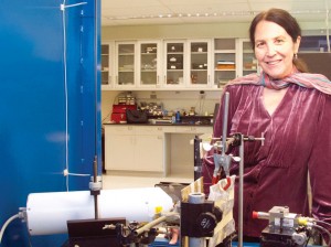
UAlbany physics professor Carolyn MacDonald of the Center for X-ray Optics, has patented a new imaging system that can help radiologists tell the difference between cancer and healthy tissue. This advance works to remedy concerns in diagnostic medicine concerning radiation dose, image contrast and resolution, and cost. Altogether, this could result in fewer breast biopsies, reduce unnecessary patient trauma and lower healthcare costs for patients and providers.
Dr. MacDonald and the University at Albany will continue to work with its expert faculty and students in order to continue fostering a Healthier New York.


